
The viewer
Diagnostic imaging. Without leaving the browser.
Radiologists open a study in Chrome, Edge, Firefox or Safari and have the tools of a reading station in front of them — on a reporting workstation, a ward PC or a laptop on call, with nothing installed on any of them.
- No client install
- No plug-ins, no Java
- WebGL2 for 3D where available
- 90+ tools
See it
The viewer, on real studies
Every capture below is the shipping viewer reading an archived study — no mock-ups, no compositing.
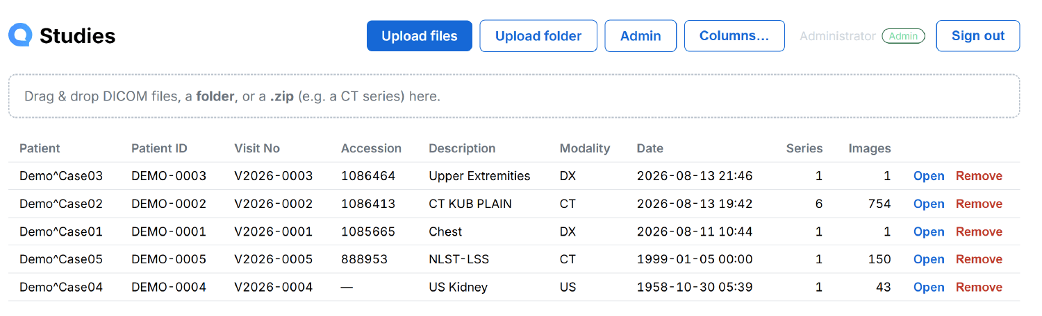
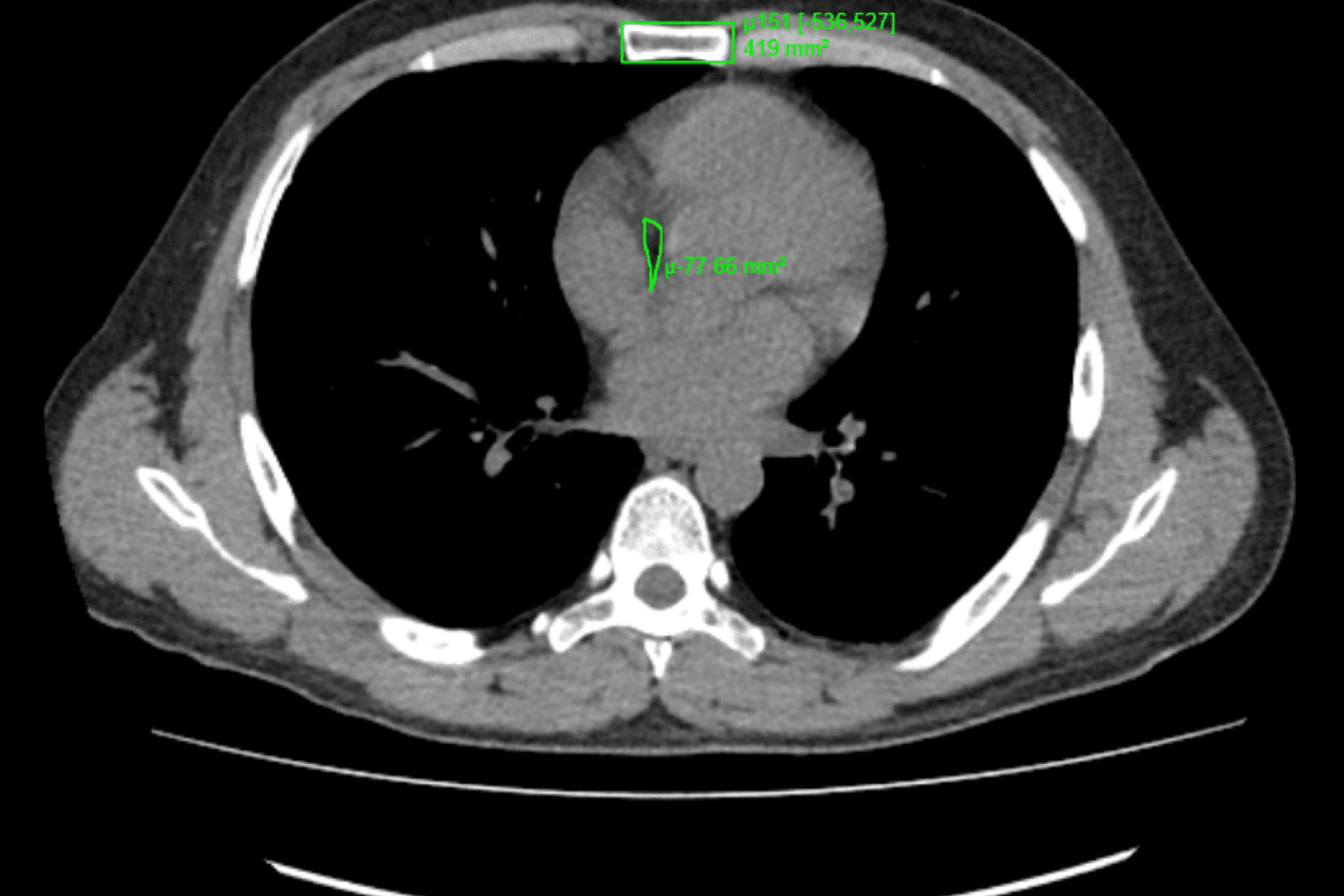
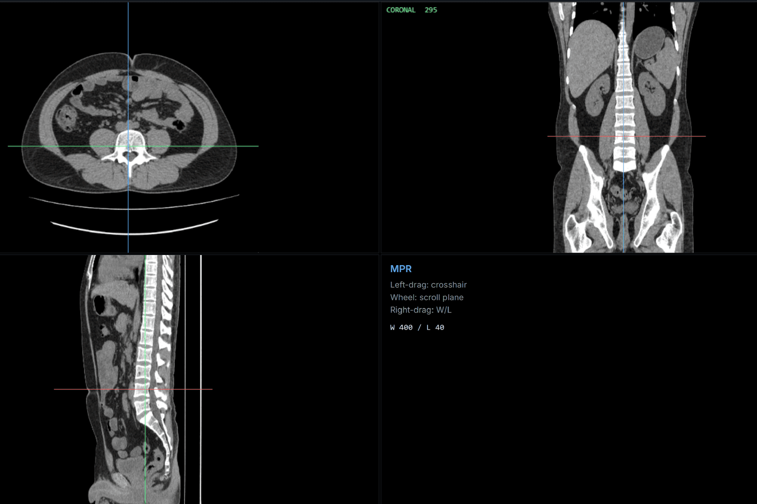
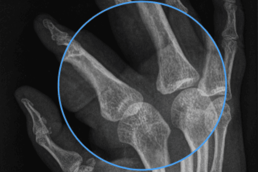
The qPACS worklist. Studies appear here seconds after acquisition, matched to their order by accession number, with patient, visit, modality, series and image counts — and drag-and-drop upload for anything arriving outside DICOM.
The toolset
Everything a reading station has
Grouped the way a radiologist reaches for them, not the way they are implemented.
Image manipulation
- Window / level — brightness & contrast
- Window presets — lung, bone, brain, soft tissue, abdomen
- Zoom, pan and scroll / stack navigation
- Fit to window, actual size 1:1, reset view
- Rotate left / right 90°, flip horizontal / vertical
- Invert greyscale
- Magnifier
Measurement & annotation
- Length, angle and Cobb angle
- Rectangle and ellipse ROI — mean, min, max, area
- Freehand ROI
- Probe — pixel value / CT Hounsfield units
- Arrow marker and text annotation
- Freehand drawing
- Eraser, delete annotation, clear all
Navigation & layout
- Cine playback with adjustable speed (FPS)
- Previous / next image and previous / next series
- Series thumbnail browser
- Layouts — 1×1, 1×2, 2×2, 3×3
- Full screen and maximise viewport
- Viewport synchronisation
- Crosshair and reference lines
Display & output
- Image overlay — patient / image data
- DICOM tag inspector
- Key image marking
- Snapshot and print
- Export image — PNG / JPEG
- Download DICOM — permission-gated
- Web upload — files, folder or .zip
Advanced visualisation
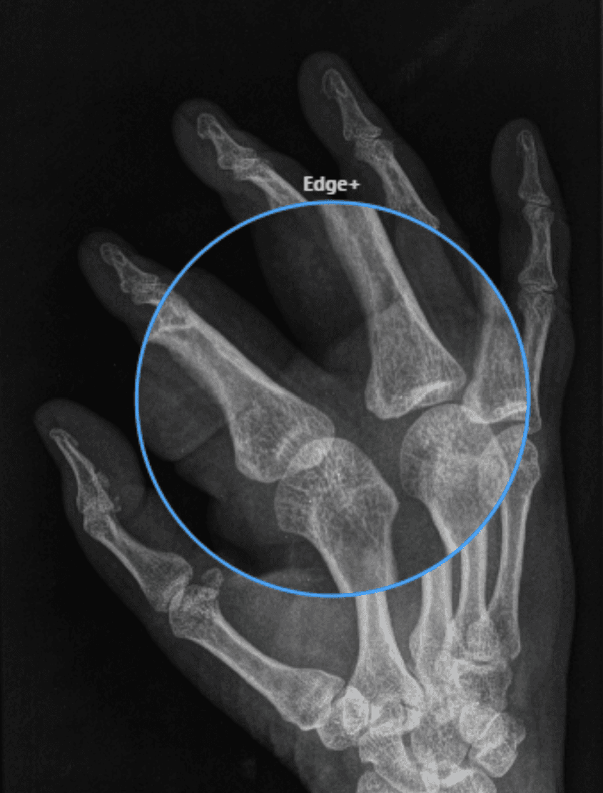
- qLens™ — local enhancement lens
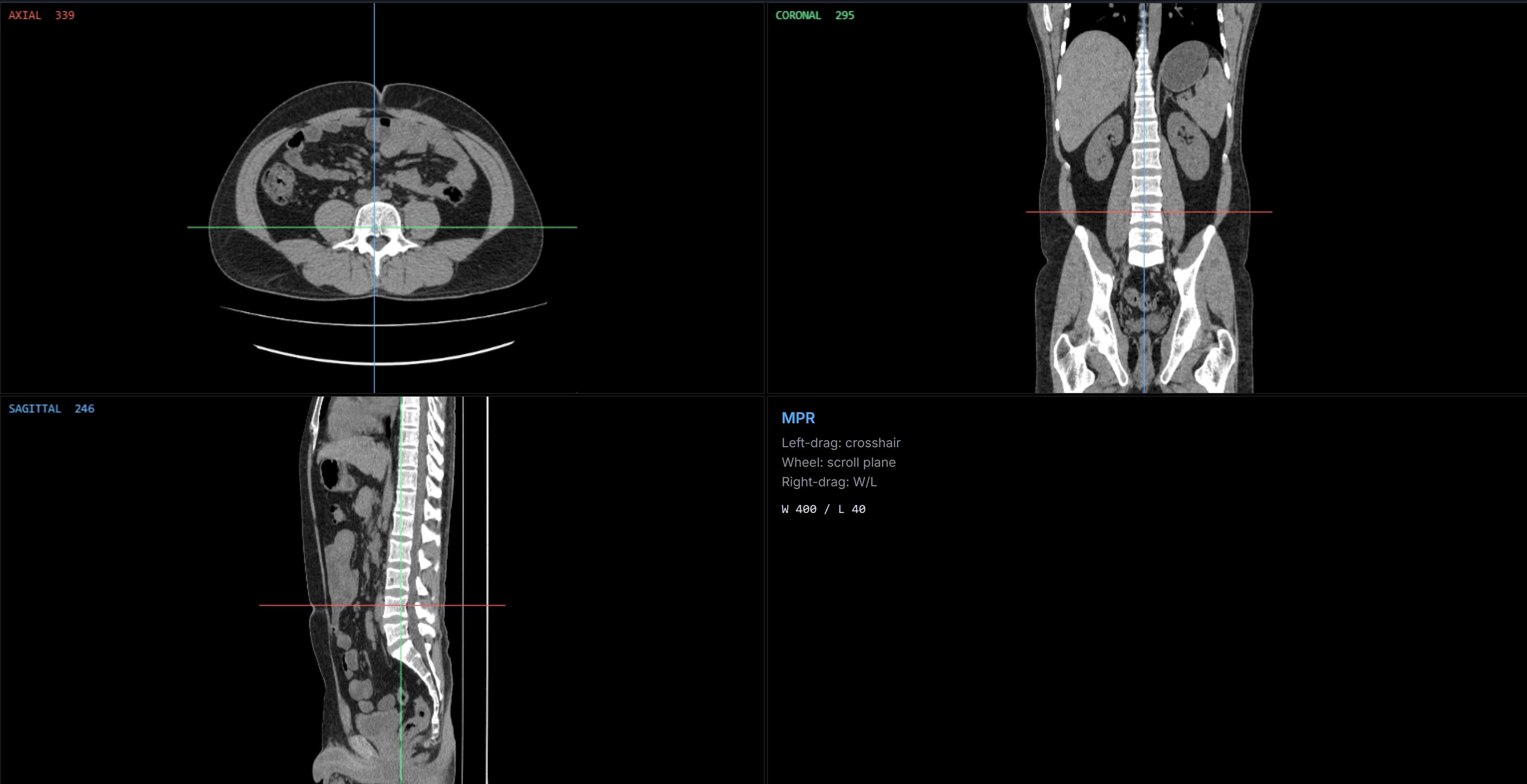
- MPR — axial, coronal, sagittal
- Linked crosshair across planes
- Per-plane scrolling
- MIP / MinIP — maximum / minimum intensity
- Average thick-slab projection
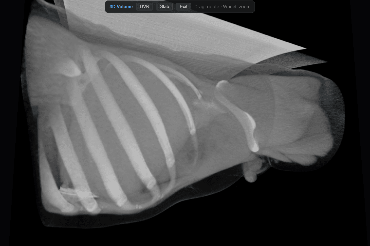
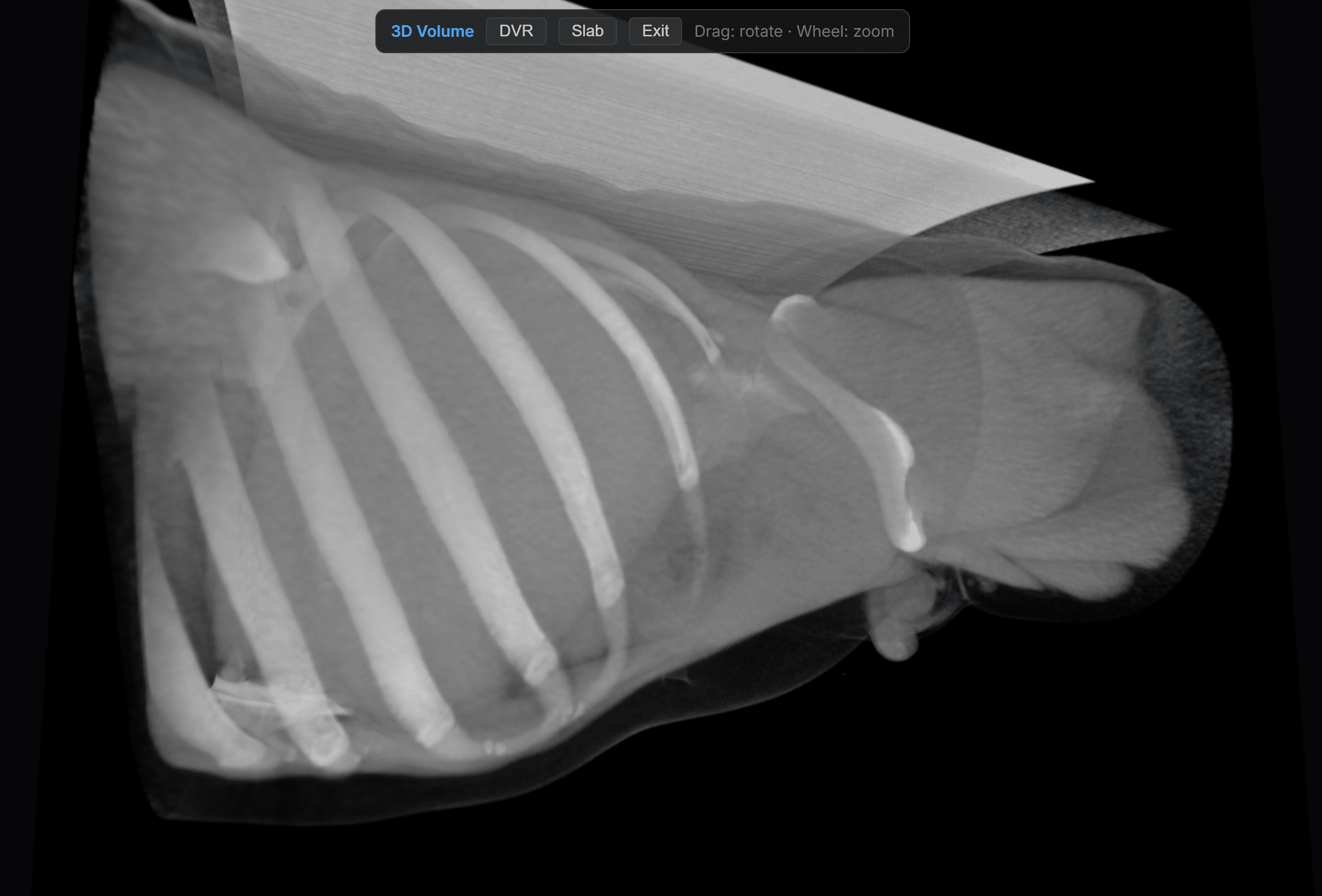
- 3D volume rendering — orbit and zoom
Performance & personalisation
- Progressive image loading and slice prefetch
- Secure caching
- Large CT / MR study handling
- Server-side decoding on retrieve
- Keyboard shortcuts
- Configurable mouse bindings
- User preferences — layout, overlays
- Hanging protocols
Innovation
qLens™ — enhancement
that never touches the study.
Edge enhancement, adaptive contrast and denoise applied inside the lens region only. The surrounding image stays exactly as acquired, and the source pixels are never modified.
What the lens can apply
Why it matters clinically
- The source DICOM pixel data is never modified — enhancement is display-only and reversible
- One-click reset returns the image to its acquired presentation
- The viewer indicates clearly when post-processing is active
- No export, no second dataset, no version of the study to reconcile later
Volume rendering
MPR, MIP and 3D in the same session
Advanced visualisation is normally a separate purchase: another workstation, another licence, another queue, and a study that has to be exported to reach it. In qPACS it is part of the same browser tab the study is already open in.
Engineered for the read, not for the demo
- Compressed studies are decoded server-side — the browser stays light
- Slices stream progressively so the first image is up immediately
- Large CT and MR series scroll without stalling
- The same interface on every PC in the building
Image handling
- Multi-frame DICOM
- Ultrasound RGB/YBR colour and cine loops
- MONOCHROME1 / MONOCHROME2
- Compressed transfer syntaxes — transcoded on retrieve
- Rescale slope / intercept and modality LUT
qPACS Mobile — Android & iOS
- Study worklist with paging and instant search
- Search by patient name, ID/MRN, accession, modality or date
- Opens studies in the qPACS viewer, with the same tools and presets
- Signs in with the same PACS account, role and permissions
- No images, reports or patient data stored on the device
- Connects to your server by address — no cloud routing
- Phone and tablet, portrait or landscape
Request a demo
See it read your studies.
The fastest way to evaluate qPACS is to point a modality at it. We install a full working system, connect it to your equipment and walk your team through a live read — on your images, on your network.
- 01Connect a modality
- 02Send a study
- 03Read it in the browser